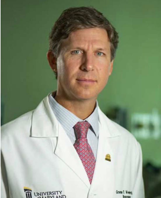
Meet the Team: Graeme Woodworth, MD
Top neurosurgeon and innovative brain cancer researcher
To hear Graeme Woodworth, MD talk to MissionGBM about his work with brain cancer patients and research, please click here.
When we first met Graeme Woodworth, MD (Chair of Neurosurgery; Director of the Brain Tumor Program, University of Maryland) in Feb-2023, we knew right away that we had to build a strong relationship with him. Top talent just has a way of creating a good first impression. Over the past 15 months, Graeme has been a frequent thought partner and a wonderful resource for many MissionGBM cases, especially the most challenging ones. We cannot thank him enough.
Dr. Woodworth’s day job is that of a practicing neurosurgeon. As such, he sees a lot of brain tumor cases for which he and his team must carefully plan and execute delicate surgeries in a manner that removes as much tumor as possible while also preserving brain executive function. No one wants to clear PACU with aphasia, memory loss, neuromotor or significant cognitive deficits.
Neurosurgery for Brain Tumors: Best to be a Careful and Unhurried Customer
Of the roughly 4000 Board-certified neurosurgeons in the US (see here and here), there are probably only about 250 that we would want operating on us. Why? Because we see a lot of MissionGBM cases in which the initial brain tumor resection was inadequately planned or executed resulting in post-surgical complications and problematic neurological deficits (usually spine surgeons in community hospitals, who may do only a few craniotomies and brain tumor resections each year; however, we have seen occasional poor outcomes from major academic medical centers). It is heartbreaking to review a case in which the patient never has a fighting chance to prosper during his/her brain cancer Journey due to poor neurosurgical intervention at the outset of the case.
We tell MissionGBM families repeatedly that they often have time to research and select a top neurosurgeon for their case instead of merely signing the consent form offered by the first neurosurgeon to show up in the ED.
It can be hard to listen to such advice when a distraught patient and family are staring at an obvious MRI hyperintensity on the ED video display (aka the “Peek-and-Shriek” moment), and you hear the words, “It looks like a brain tumor”. We know; we have been there ourselves.
While we wanted Julie’s tumor resected yesterday when she presented in the ED of a local community hospital with rapid onset aphasia, we decided to stabilize her and then transfer her to a skilled hospital of neuroscience for the craniotomy and tumor resection. Her operation occurred a week later after detailed planning was completed by her neurosurgical team (see here).
Bottom Line: You want a top neurosurgeon like Graeme Woodworth, MD working your brain tumor case. Take your time. Make an informed choice.
Innovation in Neurosurgery
Like all neurosurgeons, Dr. Woodworth knows that there are significant limitations to the adage “nothing heals like cold steel” (or “hot lasers”, if you prefer). Contemporary imaging and surgical visualization technologies simply do have enough resolution to “see” every cancer cell in the operative field. Moreover, the tumor often has invaded areas of the brain that cannot be safely resected. Finally, any surgical procedure carries multiple levels of risk, which requires that the neurosurgical team carefully plan the operation and also have contingency plans to rapidly address the spectrum of potential complications that can arise.
To address some of the limitations of brain tumor resection, Dr. Woodworth has established neurosurgical programs that extend current MR-guided tractography techniques into the realm of state-of-the-art “Connectomics” mapping (see here and here). Using these technologies allows the neurosurgical team to understand and track the critical neural circuits that define brain executive function in both the surgical planning as well as intra-operative settings. Performing neurosurgery in the absence of such neural mapping could result in cerebrovascular complications, or utterance of the infamous punchline, “Oops, there go the piano lessons!”
Dr. Woodworth is also active in the area of neurosurgical research regarding the use of Laser Interstitial Thermal Therapy (LITT; see here) in combination with other stereotactic methods to improve outcomes in brain tumor resections. Given the thermal cytoreductive nature of LITT, there is significant risk that the patient will experience post-operative cerebral edema as a result of laser-induced thermal injury. By combining MR-guided Connectomics with LITT, Dr. Woodworth’s team is able to create a de facto “awake LITT” procedure that is designed to ablate the tumor tissue with minimal thermal damage to the surrounding healthy neuronal tissue. Going further, Dr. Woodworth and colleagues are currently exploring the combined use of LITT + Proton Beam Therapy to improve clinical outcomes for both ndGBM and rGBM patients (see clinical trials NCT04699773 and NCT04181684).
Transiently Opening the BBB to Enable Better Drug Delivery
Perhaps, one of Dr. Woodworth’s most intense interests is understanding the science behind the BBB, and developing techniques to permit enhanced drug delivery across the BBB. He is a leader in the growing community of researchers focused on the application of Focused Ultrasound (FUS; see here) with microbubbles to transiently open the BBB so that therapeutically meaningful quantities of active pharmaceutical agents can gain access to the site of a brain tumor (see here, here, and here). Under normal circumstances, the intact BBB serves as stiff barrier to the transport of therapeutic agents into the brain (see here). In brain tumor cases, the BBB can act as an impediment to drug delivery into the tumor bed, thus limiting the efficacy of the treatment due to delivery of sub-therapeutic doses into the TME.
In order to treat a brain tumor, it is absolutely imperative that enough therapeutic agent reaches the TME and remains resident for sufficient time to cause the desired treatment effect according to the mechanism of action of the agent. In fact, as investors in the development of brain cancer treatment options, we will quickly reject any proposal that does not spell out a detailed plan for both (i) ensuring that the therapeutic agent can be adequately delivered to the TME; and (ii) quantitatively measuring the amount of agent within the TME (see here and here).
One of the many things that attracted us to Dr. Woodworth’s research is his insistence on developing techniques to quantitatively understand and document the neuropharmacology of FUS-enabled brain tumor drug delivery. His work is differentiated from the majority of efforts in the area by an insistence on actually measuring the concentration of the drug as opposed to simply showing qualitative images of enhanced permeability of contrast agent after FUS application. This is what is known as “rigorous neuropharmacology”, and there is just no substitute for it, if one is dedicated to translating research into successful human clinical trials instead of just curing cancer in mice.
Speaking of translational research, Dr. Woodworth has advanced his programs into multiple clinical studies (see here, here and here) with some of the early efforts beginning to yield Results That Matter (see here and here).
We thank Graeme Woodworth, MD for his continued innovation in neurosurgical technique; his research into improved methods for transport of therapeutic and diagnostic agents across the BBB; and his assistance with several of the most challenging MissionGBM cases via direct patient interaction in his practice as well as peer-to-peer consulting.


